Assembly of Capsids from Hepatitis B Virus Core Protein Progresses through Highly Populated Intermediates in the Presence and Absence of RNA
Abstract
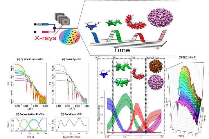
Copyright © 2020 American Chemical Society. The genetic material of viruses is protected by protein shells that are assembled from a large number of subunits in a process that is efficient and robust. Many of the mechanistic details underpinning efficient assembly of virus capsids are still unknown. The assembly mechanism of hepatitis B capsids has been intensively researched using a truncated core protein lacking the C-terminal domain responsible for binding genomic RNA. To resolve the assembly intermediates of hepatitis B virus (HBV), we studied the formation of nucleocapsids and empty capsids from full-length hepatitis B core proteins, using time-resolved small-angle X-ray scattering. We developed a detailed structural model of the HBV capsid assembly process using a combination of analysis with multivariate curve resolution, structural modeling, and Bayesian ensemble inference. The detailed structural analysis supports an assembly pathway that proceeds through the formation of two highly populated intermediates, a trimer of dimers and a partially closed shell consisting of around 40 dimers. These intermediates are on-path, transient and efficiently convert into fully formed capsids. In the presence of an RNA oligo that binds specifically to the C-terminal domain the assembly proceeds via a similar mechanism to that in the absence of nucleic acids. Comparisons between truncated and full-length HBV capsid proteins reveal that the unstructured C-terminal domain has a significant impact on the assembly process and is required to obtain a more complete mechanistic understanding of HBV capsid formation. These results also illustrate how combining scattering information from different time-points during time-resolved experiments can be utilized to derive a structural model of protein self-assembly pathways.